
Vitiligo & Pigment Cell Biology
What is vitiligo?
Vitiligo is an acquired depigmenting skin disorder characterized by the loss of melanocytes from the epidermis. It is one of common skin diseases reported to affect approximately 1% of the population worldwide, irrespective of skin color or ethnic origin.
According to the distribution of the lesions, several types of vitiligo have distinguished,
-
Localized vitiligo: basically involves one or more skin lesions in one area
- Focal vitiligo,
- Vitiligo segmentalis
-
Generalized vitiligo: involves several skin lesions in several areas
- Acrofacial vitiligo,
- Vitiligo vulgaris
- Vitiligo universalis
What is the cause of vitiligo?
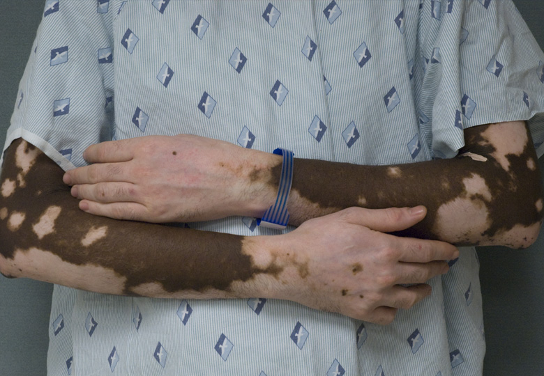 |
| Arms (Zoom In) |
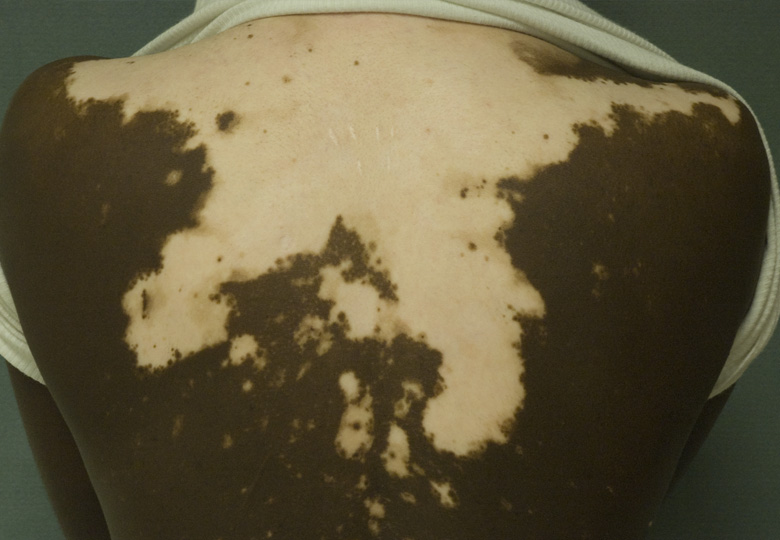 |
| Upper Back (Zoom In) |
Heredity is regarded as one of factor because some patients have familial members with vitiligo. However, the majority of patients are sporadic and non-familial cases. One hypothesis considers vitiligo an autoimmune disease. Appearance of melanocyte-specific cytotoxic T-cells and detection of autoantibody to melanocyte in the circulation indicate the involvement of both cellular and humoral immunity to the pathogenesis of vitiligo. Although the exact cause of vitiligo is still not fully understood and under investigating, it seems to be dependent on the interaction of genetic, immunologic, neurogenic, and environmental factors.
How do physicians make a diagnosis of vitiligo?
A Diagnosis of vitiligo is usually made by a primary care physician or a dermatologist according to the typical clinical features and exception of other diseases that cause leucoderma (listed below). Wood's light (a hand-held ultraviolet irradiation device) is likely used to identify areas of depigmentation. In the case of atypical presentation, skin biopsy might be taken in order to make a diagnosis.
What examinations will be recommended if I am diagnosed as vitiligo?
Patients with vitiligo often develop other autoimmune diseases and familial histories of autoimmune diseases also can be seen. Especially co-existence of vitiligo and autoimmune thyroid diseases are well documented, suggesting in adults with vitiligo, blood test for thyroid function and antinuclear antibodies are recommended.
What is the treatment of vitiligo?
The options of treatment of vitiligo are very wide ranges, including topical, photo, surgical therapies, and depigmentation. We can also choose cosmetical reagent and complementary therapies.
- Non-treatment Option: Patients whose skin type is categorized into skin type I and II can select non-treatment option after discussion — camouflage cosmetics, sunscreens
- Topical Therapy: Applied for localized vitiligo — corticosteroid, calcineurin inhibitors (tacrolimus, pimecrolimus), Vitamin D derivates, Prostaglandin E2, Antioxidants
- Phototherapy: Applied for localized vitiligo — TUVA/PUVA, Broadband UVB and Narrowband UVB
UVA = ultraviolet A, UVB = ultraviolet B - Surgical treatments: Applied for vitiligo legions stable at least from 2 years and refractory to medical treatments — autologous skin grafts, transfer of suction blisters
- Depigmentation: applied for Generalized or Vitiligo universalis that involves more than 50% of body surface area — 20% monobenzyl ether of hydroquinone, 4-Methoxyphenol, Q-switched ruby laser, cryotherapy
Appropriate treatments are chosen mainly by distribution of the lesions, duration of the disease, previous treatment, and patient's skin type. Age of patient also should be considered, because there are no studies of surgical and depigmentation treatment for children.
What is skin type?
Skin type was proposed by Fitzpatrick in 1975 according to their skin color and burning and tuning responses to sun exposure. This classification highly corresponded to the risk of skin cancer. Vitiligo is more visible in skin types IV, V, and VI, but can occur in all types of skin. Depigmented skin can sunburn more easily and might than be at high risk for skin cancer.
| Skin type | Typical features | Burning and Tanning tendency |
|---|---|---|
| I | Pale white skin, blue/hazel eyes, blond/red hair | Always burns, does not tan |
| II | Fair skin, blue eyes | Burns easily, tans poorly |
| III | Darker white skin | Tans after initial burn |
| IV | Light brown skin | Burns minimally, tans easily |
| V | Brown skin | Rarely burns, tans darkly easily |
| VI | Dark brown or black skin | Never burns, always tans darkly |
What are other diseases that cause leucoderma?
- Leucoderma senile: aging of melanocyte and keratinocyte
- Pityriasis vesicolor: skin infection of Malassezia species
- Postinflammatory disorders: Pityriasis alba, Chemical and physical agents, Sun exposure and burn, Lupus erythematosus, Scleroderma
- Lichen sclerosus: chronic skin disease predominantly found in the anogenital area
- Halo nevus: a pigmented nevus surrounded by a ring of depigmentation
- Cancer associated: Extramanmmary Paget's disease, Melanoma-associated leucoderma
- Others: Idiopathic guttate hypomelanosis, Leucoderma punctata, Progressive macular hypomelanosis of the trunk, nevus depigmentosus, piebaldism, incontinentia pigmenti achromians, phenylketouria, tuberous sclerosis, Waardenburg syndrome, Chediak-Higashi syndrome, Vogt-Koyanagi-Harada syndrome, leucoderma pseudosyphiliyicum
Recommended websites: